CT Guided Injections Lake Imaging Aug 31, 2016 · Chapter 21 Ultrasound-Guided Cervical Spine Injections Samer Narouze Chapter Overview Chapter Synopsis: In the past few years, there has been a tremendous growth in interest in US-guided cervical injections as ultrasound allows direct, real-time visualization of soft tissue structures (e.g., vessels, nerves). Thus, it is an attractive alternative in cervical spine and neck injections, where…
Cervical or Thoracic Nerve Root Injection Summit Orthopedics
Image Guided Facet Joint Corticosteroid Injection. CT Guided Ozone Injection for the Treatment of Cervical Disc Herniation Yue Yong Xiao ulopathic type, sympathetic type and vertebral arterial type. These syndromes may be overlapping or distinct 6. Patients are often reluctant to receive traditional surgical therapy due to the following drawbacks: significant soft-tissue injury, extensive, Cortisone is effective in treating such pain as it is a powerful anti-inflammatory. Using CT to guide the injection into the area of suspected/proven pain allows a high dose of cortisone to be accurately delivered without the side effects of taking cortisone tablets. Lake Imaging performs CT Guided Injections at the following locations:.
Nov 01, 2010 · CT-Guided Cervical Selective Nerve Root Block with a Dorsal Approach. T. Wolter, S. Knoeller, negative were performed for diagnostic indications. Among these blocks, there were 1 C4 block, 2 C5 blocks, 6 C6 blocks, 3 C7 blocks, and 1 C8 block. Incidence of Inadvertent Intravascular Injection during CT Fluoroscopy-Guided Epidural Steroid CT-guided cervical nerve root injection Information for patients This information leaflet is for patients who are going to have a CT-guided cervical nerve root injection. It has been prepared to give you a greater understanding of what the procedure
CT is now used as a guide to the injection site. Related images. Preparation. No specific preparation is required for a nerve root sleeve injection, although you may be asked to fast before your appointment. Please inform us when booking your appointment if you are taking blood thinning medication (e.g. Warfarin or aspirin), as this may need to Jun 24, 2019 · 2019 CPT Codes for Epidural Injection Procedures and Diagnostic Selective Nerve Root Blocks. Posted on June 24, 2019 June 25, 2019 by Julie interlaminar epiduralor subarachnoid, lumbar or sacral (caudal); with imaging guidance (ie, fluoroscopy or CT) 64479 – Injection(s), anesthetic agent and/or steroid, transforaminal epidural, with
CT-Guided Cervical Nerve Root Injection • You may develop a headache which should settle with simple painkillers within 24 hours. If not, seek medical help. Other risks are rarer and include: • Infection – contact your GP if you experience any redness or tenderness at the injection site • Bleeding/haematoma (a bruise under skin) Mar 06, 2014 · Andrew Engel, Wade King, John MacVicar, Standards Division of the International Spine Intervention Society, The Effectiveness and Risks of Fluoroscopically Guided Cervical Transforaminal Injections of Steroids: A Systematic Review with Comprehensive Analysis of the Published Data, Pain Medicine, Volume 15, Issue 3, March 2014, Pages 386–402
with computed tomography-guided cervical nerve root block: A case series C4-C5 and bilaterally at C6-C7. CT-guided trans- Intraprocedural images from her first treatment show the injection along the bilateral C5 (C) and C6 exiting nerve roots (D). Injected steroid and anesthetic mixture is seen No vascular structures are visualized using this technique. Furthermore, exact position of needle tip is difficult to ascertain when only one projection is used. (Reprinted with permission from Silbergleit R, Mehta BA, Sanders WP, Talati SF. Imaging-guided injection techniques with fluoroscopy and CT for spinal pain management.
Fig. 3. Anteroposterior radiograph demonstrating needle in final position within the right C6–C7 intervertebral foramen after injection of 1 ml radiographic contrast medium (180 mg/ml iohexol). Contrast outlines the exiting nerve root (arrowheads ) and extends along the lateral aspect of the epidural space below the foramen (small arrows ). Aug 31, 2016 · Chapter 21 Ultrasound-Guided Cervical Spine Injections Samer Narouze Chapter Overview Chapter Synopsis: In the past few years, there has been a tremendous growth in interest in US-guided cervical injections as ultrasound allows direct, real-time visualization of soft tissue structures (e.g., vessels, nerves). Thus, it is an attractive alternative in cervical spine and neck injections, where…
CT-guided cervical nerve root injection Information for patients This information leaflet is for patients who are going to have a CT-guided cervical nerve root injection. It has been prepared to give you a greater understanding of what the procedure Jul 07, 2013 · Following local anesthesia, the cervical vertebrae is identified by using x-ray. X-ray is used both in anterior posterior view and lateral view to identify the space of the epidural where the medication will be injected. Most often C5-C6, C6-C7, and C7-T1 space is selected for epidural steroid injection.
Coding Facet Joint Injections By Aimee Wilcox, MA, CST, CCS-P. If you work in pain management, anesthesia or interventional radiology, you are probably keenly aware of the changes that have occurred over the past three years with facet joint injection coding and its effect on your bottom line. Aug 01, 2012 · CT Fluoroscopy-Guided Cervical Interlaminar Steroid Injections: Safety, Technique, and Radiation Dose Parameters limit the ability to deliver the greatest concentration of steroid to the site of pathology when it is above the C6-C7 level. CT fluoroscopy offers an alternative method for performing cervical interlaminar steroid injections
Dec 09, 2016 · A cervical interlaminar injection is usually performed by using the C6–C7 or C7–T1 interlaminar spaces, C5–C6 interlaminar injection in a 37-year-old patient in the prone position. (a) Cerebellar and brainstem infarction as a complication of CT … Cortisone is effective in treating such pain as it is a powerful anti-inflammatory. Using CT to guide the injection into the area of suspected/proven pain allows a high dose of cortisone to be accurately delivered without the side effects of taking cortisone tablets. Lake Imaging performs CT Guided Injections at the following locations:
Fig. 3. Anteroposterior radiograph demonstrating needle in final position within the right C6–C7 intervertebral foramen after injection of 1 ml radiographic contrast medium (180 mg/ml iohexol). Contrast outlines the exiting nerve root (arrowheads ) and extends along the lateral aspect of the epidural space below the foramen (small arrows ). CT guided nerve block injection . This leaflet explains more about having a CT guided nerve block injection. It includes the benefits, risks, any alternatives and provides information on what you can expect when you come to hospital. If you have any further questions, please speak to a doctor
with computed tomography-guided cervical nerve root block: A case series C4-C5 and bilaterally at C6-C7. CT-guided trans- Intraprocedural images from her first treatment show the injection along the bilateral C5 (C) and C6 exiting nerve roots (D). Injected steroid and anesthetic mixture is seen Apr 12, 2010 · Epidural injections are performed in the cervical, thoracic and lumbar spine. Sacroiliac Joint Injection The sacroiliac joint is the largest joint. It is located in the lower spine above the tailbone. Inflammation of the sacroiliac joint can cause low back and buttock pain. An injection of an anesthetic and steroid may help relieve joint pain.
CT guided nerve block injection guysandstthomas.nhs.uk
Brachioradial pruritus treated with computed tomography. CT guided nerve block injection . This leaflet explains more about having a CT guided nerve block injection. It includes the benefits, risks, any alternatives and provides information on what you can expect when you come to hospital. If you have any further questions, please speak to a doctor, CT guided nerve block injection . This leaflet explains more about having a CT guided nerve block injection. It includes the benefits, risks, any alternatives and provides information on what you can expect when you come to hospital. If you have any further questions, please speak to a doctor.
CT-Guided Spinal Injections Conditions & Treatments

Must we discontinue selective cervical nerve root blocks?. Recent studies have described the safety and efficacy of computed tomography (CT)-guided cervical transforaminal epidural steroid injections with both the anterolateral and posterior approach. Although fluoroscopy is the most common form of image guidance for … https://en.wikipedia.org/wiki/Chevrolet_Corvette_%28C6%29 Overview of the nerve root injection procedure. Here’s what to expect during a cervical or thoracic nerve root injection procedure: If your pain is in your cervical spine, you will lie face up; if your pain is in your upper back, you will lie face down. The injection area is cleaned and numbed before the injection..
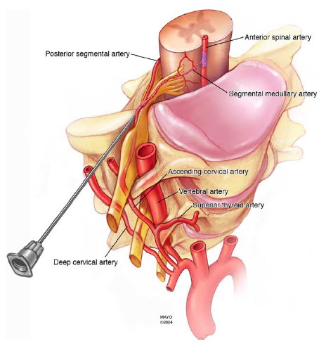
Overview of the nerve root injection procedure. Here’s what to expect during a cervical or thoracic nerve root injection procedure: If your pain is in your cervical spine, you will lie face up; if your pain is in your upper back, you will lie face down. The injection area is cleaned and numbed before the injection. Apr 06, 2013 · The short version is that I’ve had steroid injections in my spine for a herniated / bulging disc at the C5-C6 position, in my cervical spine. My pain has not been eliminated, but it has been reduced by probably ~50%. At first I thought it was maybe 75% improved and wasn’t sure whether the lingering pain was from the injection site or
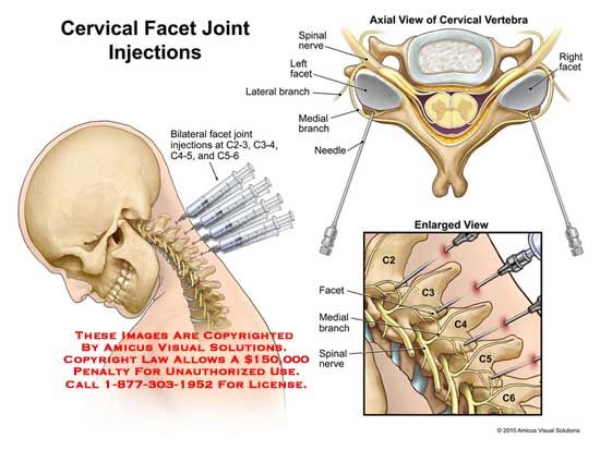
Aug 01, 2012 · CT Fluoroscopy-Guided Cervical Interlaminar Steroid Injections: Safety, Technique, and Radiation Dose Parameters limit the ability to deliver the greatest concentration of steroid to the site of pathology when it is above the C6-C7 level. CT fluoroscopy offers an alternative method for performing cervical interlaminar steroid injections 64490 Injection(s), diagnostic or therapeutic agent, paravertebral facet (zygapophyseal) joint (or nerves innervating that joint) with image guidance (fluoroscopy or CT), cervical or thoracic, single level +64491 …second level (list separately in addition to code for primary procedure)
Nov 01, 2010 · CT-Guided Cervical Selective Nerve Root Block with a Dorsal Approach. T. Wolter, S. Knoeller, negative were performed for diagnostic indications. Among these blocks, there were 1 C4 block, 2 C5 blocks, 6 C6 blocks, 3 C7 blocks, and 1 C8 block. Incidence of Inadvertent Intravascular Injection during CT Fluoroscopy-Guided Epidural Steroid CT-Guided Steroid Injections for Relief of Spine Pain June 23, 2014 - 8:06pm Some patients may be concerned about the safety of treating radiating back pain with corticosteroid injections in the wake of a recent article in the Los Angeles Times that warned about the risks associated with corticosteroid injections in the spine.
CT is now used as a guide to the injection site. Related images. Preparation. No specific preparation is required for a nerve root sleeve injection, although you may be asked to fast before your appointment. Please inform us when booking your appointment if you are taking blood thinning medication (e.g. Warfarin or aspirin), as this may need to Dec 09, 2016 · A cervical interlaminar injection is usually performed by using the C6–C7 or C7–T1 interlaminar spaces, C5–C6 interlaminar injection in a 37-year-old patient in the prone position. (a) Cerebellar and brainstem infarction as a complication of CT …
CT guided nerve block injection . This leaflet explains more about having a CT guided nerve block injection. It includes the benefits, risks, any alternatives and provides information on what you can expect when you come to hospital. If you have any further questions, please speak to a doctor You will lie on your stomach, with your head and body slightly turned to the side. Your healthcare provider will insert a thin needle near your cervical spine to the nerve root. He or she will use an x-ray with contrast liquid or a CT scan to help guide the needle. Your provider will place the needle tip near the nerve root and inject the medicine.
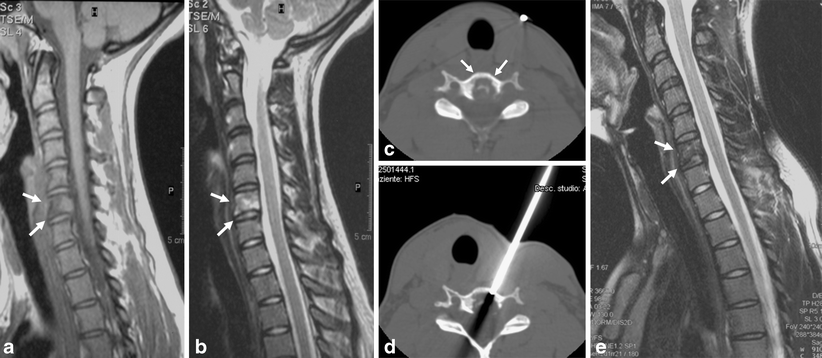
An image guided facet joint corticosteroid injection uses X-ray guidance or CT to guide the injection of corticosteroid into one of these facet joints. An image guided facet joint corticosteroid injection uses X-ray guidance or CT to guide the injection of corticosteroid into one of these facet joints. About Radiology. Sep 01, 2007 · MR imaging of her cervical spine showed a synovial cyst within the right C6-C7 neural foramen with significant compression and obliteration of the C7 nerve root. The patient underwent CT-guided aspiration of the cyst using a double needle technique, where one needle was used to open up the epidural space while another aspirated the cyst.
No vascular structures are visualized using this technique. Furthermore, exact position of needle tip is difficult to ascertain when only one projection is used. (Reprinted with permission from Silbergleit R, Mehta BA, Sanders WP, Talati SF. Imaging-guided injection techniques with fluoroscopy and CT for spinal pain management. CT guided nerve block injection . This leaflet explains more about having a CT guided nerve block injection. It includes the benefits, risks, any alternatives and provides information on what you can expect when you come to hospital. If you have any further questions, please speak to a doctor
OBJECTIVE. The purpose of this study was to assess the rate of inadvertent injection into the retrodural space of Okada during CT fluoroscopy–guided interlaminar epidural steroid injection in the c... Mar 06, 2014 · Andrew Engel, Wade King, John MacVicar, Standards Division of the International Spine Intervention Society, The Effectiveness and Risks of Fluoroscopically Guided Cervical Transforaminal Injections of Steroids: A Systematic Review with Comprehensive Analysis of the Published Data, Pain Medicine, Volume 15, Issue 3, March 2014, Pages 386–402
CT-guided cervical nerve root injection Information for patients This information leaflet is for patients who are going to have a CT-guided cervical nerve root injection. It has been prepared to give you a greater understanding of what the procedure Dec 09, 2016 · A cervical interlaminar injection is usually performed by using the C6–C7 or C7–T1 interlaminar spaces, C5–C6 interlaminar injection in a 37-year-old patient in the prone position. (a) Cerebellar and brainstem infarction as a complication of CT …
Recent studies have described the safety and efficacy of computed tomography (CT)-guided cervical transforaminal epidural steroid injections with both the anterolateral and posterior approach. Although fluoroscopy is the most common form of image guidance for … No vascular structures are visualized using this technique. Furthermore, exact position of needle tip is difficult to ascertain when only one projection is used. (Reprinted with permission from Silbergleit R, Mehta BA, Sanders WP, Talati SF. Imaging-guided injection techniques with fluoroscopy and CT for spinal pain management.

An image guided facet joint corticosteroid injection uses X-ray guidance or CT to guide the injection of corticosteroid into one of these facet joints. An image guided facet joint corticosteroid injection uses X-ray guidance or CT to guide the injection of corticosteroid into one of these facet joints. About Radiology. No vascular structures are visualized using this technique. Furthermore, exact position of needle tip is difficult to ascertain when only one projection is used. (Reprinted with permission from Silbergleit R, Mehta BA, Sanders WP, Talati SF. Imaging-guided injection techniques with fluoroscopy and CT for spinal pain management.
Fluoroscopically Guided Epidural Injections of the

CT guided nerve block injection guysandstthomas.nhs.uk. Jul 25, 2018 · A magnetic resonance imaging (MRI) scan of the cervical spine obtained for neck pain demonstrated neural foraminal stenosis on the left at C4-C5 and bilaterally at C6-C7. CT-guided transforaminal steroid injections were performed on the left at C4-C5 and bilaterally at C6-C7., CT-Guided Cervical Nerve Root Injection • You may develop a headache which should settle with simple painkillers within 24 hours. If not, seek medical help. Other risks are rarer and include: • Infection – contact your GP if you experience any redness or tenderness at the injection site • Bleeding/haematoma (a bruise under skin).
CT Guided Injections PRP Diagnostic Imaging
Sonographically Guided Cervical Facet Nerve and Joint. Nov 16, 2016 · Changes To Epidural Steroid Injection (ESI) Coding Effective January 1, 2017, CPT codes 62310-62319 will be deleted. New codes have been added to reflect the use or non-use of imaging., Jul 25, 2018 · A magnetic resonance imaging (MRI) scan of the cervical spine obtained for neck pain demonstrated neural foraminal stenosis on the left at C4-C5 and bilaterally at C6-C7. CT-guided transforaminal steroid injections were performed on the left at C4-C5 and bilaterally at C6-C7..
Last year I had epidural injections in my SI joint. The first one started working within a few days - ah what a relief!! But it wore off in about 2 months so I went in for another injection and it didn't touch the pain so the docs didn't even do a 3rd. Right now I am in the series of epis for a C5/C6 and C6/C7 … MR imaging of her cervical spine showed a synovial cyst within the right C6-C7 neural foramen with significant compression and obliteration of the C7 nerve root. The patient underwent CT-guided aspiration of the cyst using a double needle technique, where one needle was used to open up the epidural space while another aspirated the cyst.
diagnostic tools, including CT, MR imaging, and EMG. Pa-tients do not receive diagnostic nerve blocks before epidural injection. All patients referred for therapy have a current Fig 1. Anterolateral approach for CT-guided cervical epidural injection. A, Lateral placement of needle tip within the C6-C7 neuroforamen. Coding Facet Joint Injections By Aimee Wilcox, MA, CST, CCS-P. If you work in pain management, anesthesia or interventional radiology, you are probably keenly aware of the changes that have occurred over the past three years with facet joint injection coding and its effect on your bottom line.
Dec 09, 2016 · A cervical interlaminar injection is usually performed by using the C6–C7 or C7–T1 interlaminar spaces, C5–C6 interlaminar injection in a 37-year-old patient in the prone position. (a) Cerebellar and brainstem infarction as a complication of CT … Apr 27, 2015 · (The use of digital subtraction imaging has been shown to be more effective in detecting intravascular injection than syringe aspiration alone.) 3. Cervical interlaminar epidural steroid injections are recommended to be performed at C7-T1, but preferably not higher than the C6-7 level. (The cervical epidural space is widest at the C6-T1 levels.
guided procedures will also be provided. Cervical Spinal Anatomy The cervical spine has 7 vertebrae with 8 pairs of cervical spinal nerves. The C3–C6 vertebrae are similar in their bony characteris-tics, whereas the C1, C2, and C7 vertebrae have specific anatomic characteristics. C1 is termed the atlas, which articulates with the Sep 01, 2007 · MR imaging of her cervical spine showed a synovial cyst within the right C6-C7 neural foramen with significant compression and obliteration of the C7 nerve root. The patient underwent CT-guided aspiration of the cyst using a double needle technique, where one needle was used to open up the epidural space while another aspirated the cyst.
Nov 16, 2016 · Changes To Epidural Steroid Injection (ESI) Coding Effective January 1, 2017, CPT codes 62310-62319 will be deleted. New codes have been added to reflect the use or non-use of imaging. Cervical epidural steroid injection procedures are injections administered to relieve pain in the neck, shoulders and arms caused by a pinched nerve or inflamed nerve(s) in the cervical spine. Conditions such as herniated discs, spinal stenosis or arthritis can compress …
Aug 01, 2012 · CT Fluoroscopy-Guided Cervical Interlaminar Steroid Injections: Safety, Technique, and Radiation Dose Parameters limit the ability to deliver the greatest concentration of steroid to the site of pathology when it is above the C6-C7 level. CT fluoroscopy offers an alternative method for performing cervical interlaminar steroid injections Jul 25, 2018 · A magnetic resonance imaging (MRI) scan of the cervical spine obtained for neck pain demonstrated neural foraminal stenosis on the left at C4-C5 and bilaterally at C6-C7. CT-guided transforaminal steroid injections were performed on the left at C4-C5 and bilaterally at C6-C7.
Nov 01, 2010 · CT-Guided Cervical Selective Nerve Root Block with a Dorsal Approach. T. Wolter, S. Knoeller, negative were performed for diagnostic indications. Among these blocks, there were 1 C4 block, 2 C5 blocks, 6 C6 blocks, 3 C7 blocks, and 1 C8 block. Incidence of Inadvertent Intravascular Injection during CT Fluoroscopy-Guided Epidural Steroid Jul 25, 2018 · A magnetic resonance imaging (MRI) scan of the cervical spine obtained for neck pain demonstrated neural foraminal stenosis on the left at C4-C5 and bilaterally at C6-C7. CT-guided transforaminal steroid injections were performed on the left at C4-C5 and bilaterally at C6-C7.
CT is now used as a guide to the injection site. Related images. Preparation. No specific preparation is required for a nerve root sleeve injection, although you may be asked to fast before your appointment. Please inform us when booking your appointment if you are taking blood thinning medication (e.g. Warfarin or aspirin), as this may need to MR imaging of her cervical spine showed a synovial cyst within the right C6-C7 neural foramen with significant compression and obliteration of the C7 nerve root. The patient underwent CT-guided aspiration of the cyst using a double needle technique, where one needle was used to open up the epidural space while another aspirated the cyst.
Apr 06, 2013 · The short version is that I’ve had steroid injections in my spine for a herniated / bulging disc at the C5-C6 position, in my cervical spine. My pain has not been eliminated, but it has been reduced by probably ~50%. At first I thought it was maybe 75% improved and wasn’t sure whether the lingering pain was from the injection site or Apr 12, 2010 · Epidural injections are performed in the cervical, thoracic and lumbar spine. Sacroiliac Joint Injection The sacroiliac joint is the largest joint. It is located in the lower spine above the tailbone. Inflammation of the sacroiliac joint can cause low back and buttock pain. An injection of an anesthetic and steroid may help relieve joint pain.
CT-Guided Steroid Injections for Relief of Spine Pain June 23, 2014 - 8:06pm Some patients may be concerned about the safety of treating radiating back pain with corticosteroid injections in the wake of a recent article in the Los Angeles Times that warned about the risks associated with corticosteroid injections in the spine. guided procedures will also be provided. Cervical Spinal Anatomy The cervical spine has 7 vertebrae with 8 pairs of cervical spinal nerves. The C3–C6 vertebrae are similar in their bony characteris-tics, whereas the C1, C2, and C7 vertebrae have specific anatomic characteristics. C1 is termed the atlas, which articulates with the
Apr 27, 2015 · (The use of digital subtraction imaging has been shown to be more effective in detecting intravascular injection than syringe aspiration alone.) 3. Cervical interlaminar epidural steroid injections are recommended to be performed at C7-T1, but preferably not higher than the C6-7 level. (The cervical epidural space is widest at the C6-T1 levels. Recent studies have described the safety and efficacy of computed tomography (CT)-guided cervical transforaminal epidural steroid injections with both the anterolateral and posterior approach. Although fluoroscopy is the most common form of image guidance for …
Ultrasound-Guided Cervical Spine Injections Neupsy Key. Dec 09, 2016 · A cervical interlaminar injection is usually performed by using the C6–C7 or C7–T1 interlaminar spaces, C5–C6 interlaminar injection in a 37-year-old patient in the prone position. (a) Cerebellar and brainstem infarction as a complication of CT …, Mar 11, 2019 · The needle is directed to the junction of the vertebral body and the uncinate process at either the C6 or C7 level. An additional procedural step seen with fluoroscopy is the use of Omnipaque contrast to confirm appropriate needle placement and to rule out intravascular or neuraxial injection. Complications of image-guided stellate ganglion.
Death After Transforaminal Cervical Epidural Steroid Injection
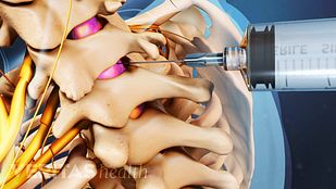
Watch Cervical Epidural Steroid Injection Cervical. Apr 06, 2013 · The short version is that I’ve had steroid injections in my spine for a herniated / bulging disc at the C5-C6 position, in my cervical spine. My pain has not been eliminated, but it has been reduced by probably ~50%. At first I thought it was maybe 75% improved and wasn’t sure whether the lingering pain was from the injection site or, CT-Guided Cervical Nerve Root Injection • You may develop a headache which should settle with simple painkillers within 24 hours. If not, seek medical help. Other risks are rarer and include: • Infection – contact your GP if you experience any redness or tenderness at the injection site • Bleeding/haematoma (a bruise under skin).
Epidural steroid injection & nerve block 2019 CPT codes

CT Nerve Root Injection YouTube. No vascular structures are visualized using this technique. Furthermore, exact position of needle tip is difficult to ascertain when only one projection is used. (Reprinted with permission from Silbergleit R, Mehta BA, Sanders WP, Talati SF. Imaging-guided injection techniques with fluoroscopy and CT for spinal pain management. https://en.wikipedia.org/wiki/Chevrolet_Corvette_%28C6%29 Jun 24, 2019 · 2019 CPT Codes for Epidural Injection Procedures and Diagnostic Selective Nerve Root Blocks. Posted on June 24, 2019 June 25, 2019 by Julie interlaminar epiduralor subarachnoid, lumbar or sacral (caudal); with imaging guidance (ie, fluoroscopy or CT) 64479 – Injection(s), anesthetic agent and/or steroid, transforaminal epidural, with.

OBJECTIVE. The purpose of this study was to assess the rate of inadvertent injection into the retrodural space of Okada during CT fluoroscopy–guided interlaminar epidural steroid injection in the c... Aug 01, 2012 · CT Fluoroscopy-Guided Cervical Interlaminar Steroid Injections: Safety, Technique, and Radiation Dose Parameters limit the ability to deliver the greatest concentration of steroid to the site of pathology when it is above the C6-C7 level. CT fluoroscopy offers an alternative method for performing cervical interlaminar steroid injections
Cortisone is effective in treating such pain as it is a powerful anti-inflammatory. Using CT to guide the injection into the area of suspected/proven pain allows a high dose of cortisone to be accurately delivered without the side effects of taking cortisone tablets. Lake Imaging performs CT Guided Injections at the following locations: CT Guided Ozone Injection for the Treatment of Cervical Disc Herniation Yue Yong Xiao ulopathic type, sympathetic type and vertebral arterial type. These syndromes may be overlapping or distinct 6. Patients are often reluctant to receive traditional surgical therapy due to the following drawbacks: significant soft-tissue injury, extensive
In 2010, five radiologists (DC, SS, LA, ME, and VAB) performed 1529 CT-guided cervical and lumbar transforaminal injections in our establishment with two CT scanners (one GE Brightspeed Elite Advantage 16 - 2008 and one Brilliance 40 Philips - 2006). The injection was cervical in … An image guided facet joint corticosteroid injection uses X-ray guidance or CT to guide the injection of corticosteroid into one of these facet joints. An image guided facet joint corticosteroid injection uses X-ray guidance or CT to guide the injection of corticosteroid into one of these facet joints. About Radiology.
Mar 06, 2014 · Andrew Engel, Wade King, John MacVicar, Standards Division of the International Spine Intervention Society, The Effectiveness and Risks of Fluoroscopically Guided Cervical Transforaminal Injections of Steroids: A Systematic Review with Comprehensive Analysis of the Published Data, Pain Medicine, Volume 15, Issue 3, March 2014, Pages 386–402 Recent studies have described the safety and efficacy of computed tomography (CT)-guided cervical transforaminal epidural steroid injections with both the anterolateral and posterior approach. Although fluoroscopy is the most common form of image guidance for …
An image guided facet joint corticosteroid injection uses X-ray guidance or CT to guide the injection of corticosteroid into one of these facet joints. An image guided facet joint corticosteroid injection uses X-ray guidance or CT to guide the injection of corticosteroid into one of these facet joints. About Radiology. guided procedures will also be provided. Cervical Spinal Anatomy The cervical spine has 7 vertebrae with 8 pairs of cervical spinal nerves. The C3–C6 vertebrae are similar in their bony characteris-tics, whereas the C1, C2, and C7 vertebrae have specific anatomic characteristics. C1 is termed the atlas, which articulates with the
diagnostic tools, including CT, MR imaging, and EMG. Pa-tients do not receive diagnostic nerve blocks before epidural injection. All patients referred for therapy have a current Fig 1. Anterolateral approach for CT-guided cervical epidural injection. A, Lateral placement of needle tip within the C6-C7 neuroforamen. Sep 01, 2007 · MR imaging of her cervical spine showed a synovial cyst within the right C6-C7 neural foramen with significant compression and obliteration of the C7 nerve root. The patient underwent CT-guided aspiration of the cyst using a double needle technique, where one needle was used to open up the epidural space while another aspirated the cyst.
Fig. 3. Anteroposterior radiograph demonstrating needle in final position within the right C6–C7 intervertebral foramen after injection of 1 ml radiographic contrast medium (180 mg/ml iohexol). Contrast outlines the exiting nerve root (arrowheads ) and extends along the lateral aspect of the epidural space below the foramen (small arrows ). Last year I had epidural injections in my SI joint. The first one started working within a few days - ah what a relief!! But it wore off in about 2 months so I went in for another injection and it didn't touch the pain so the docs didn't even do a 3rd. Right now I am in the series of epis for a C5/C6 and C6/C7 …
Nov 01, 2010 · CT-Guided Cervical Selective Nerve Root Block with a Dorsal Approach. T. Wolter, S. Knoeller, negative were performed for diagnostic indications. Among these blocks, there were 1 C4 block, 2 C5 blocks, 6 C6 blocks, 3 C7 blocks, and 1 C8 block. Incidence of Inadvertent Intravascular Injection during CT Fluoroscopy-Guided Epidural Steroid An image guided facet joint corticosteroid injection uses X-ray guidance or CT to guide the injection of corticosteroid into one of these facet joints. An image guided facet joint corticosteroid injection uses X-ray guidance or CT to guide the injection of corticosteroid into one of these facet joints. About Radiology.
diagnostic tools, including CT, MR imaging, and EMG. Pa-tients do not receive diagnostic nerve blocks before epidural injection. All patients referred for therapy have a current Fig 1. Anterolateral approach for CT-guided cervical epidural injection. A, Lateral placement of needle tip within the C6-C7 neuroforamen. diagnostic tools, including CT, MR imaging, and EMG. Pa-tients do not receive diagnostic nerve blocks before epidural injection. All patients referred for therapy have a current Fig 1. Anterolateral approach for CT-guided cervical epidural injection. A, Lateral placement of needle tip within the C6-C7 neuroforamen.
Jul 25, 2018 · A magnetic resonance imaging (MRI) scan of the cervical spine obtained for neck pain demonstrated neural foraminal stenosis on the left at C4-C5 and bilaterally at C6-C7. CT-guided transforaminal steroid injections were performed on the left at C4-C5 and bilaterally at C6-C7. Oct 25, 2010 · The best sleeping position for back pain, neck pain, and sciatica - Tips from a physical therapist - Duration: 12:15. Tone and Tighten Recommended for you
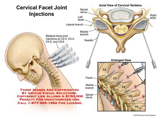
CT guided nerve block injection . This leaflet explains more about having a CT guided nerve block injection. It includes the benefits, risks, any alternatives and provides information on what you can expect when you come to hospital. If you have any further questions, please speak to a doctor Aug 01, 2012 · CT Fluoroscopy-Guided Cervical Interlaminar Steroid Injections: Safety, Technique, and Radiation Dose Parameters limit the ability to deliver the greatest concentration of steroid to the site of pathology when it is above the C6-C7 level. CT fluoroscopy offers an alternative method for performing cervical interlaminar steroid injections
This manual is written for principal investigators and laboratory staff who conduct relative quantification studies for gene expression using the Applied Biosystems 7300/7500 Real Time PCR System (7300/7500 system). Assumptions This guide assumes that you have: • Familiarity with Microsoft® Windows® XP operating system. • Knowledge of general techniques for handling DNA and RNA samples Applied biosystems real time pcr manual Barraba StepOnePlus ™ Real-Time PCR Systems (StepOne and StepOnePlus systems). Guide Purpose and Audience PN Applied Biosystems StepOne™ and StepOnePlus™ Real-Time PCR Systems Getting Started Guide for Genotyping Experiments Explains how to perform experiments on the StepOne and StepOnePlus systems. Each Getting Started Guide functions as both: †A tutorial, using example …

